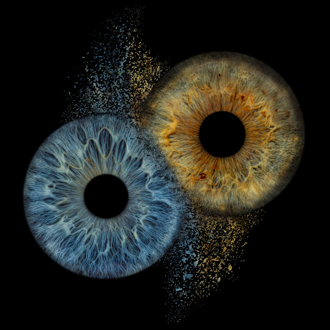
The iris is the thin, circular structure that surrounds the pupil of the eye, and it plays a critical role in regulating the amount of light that enters the eye. In this article, we will provide a scientific overview of the anatomy and function of the iris.
Anatomy of the Iris
The iris is composed of two layers of pigmented cells: the anterior pigmented epithelium and the posterior pigmented epithelium. The anterior layer is located on the outer surface of the iris and is continuous with the pigmented layer of the ciliary body. The posterior layer is located on the inner surface of the iris and is in contact with the aqueous humor. Together, these two layers form the stroma of the iris.
The stroma of the iris is composed of connective tissue and smooth muscle fibers. The connective tissue is made up of collagen and elastin fibers, which give the iris its strength and elasticity. The smooth muscle fibers are arranged in two layers: the dilator pupillae and the sphincter pupillae. The dilator pupillae muscle is responsible for enlarging the pupil, while the sphincter pupillae muscle is responsible for constricting the pupil.
Function of the Iris
The primary function of the iris is to regulate the amount of light that enters the eye. The iris accomplishes this through a process known as the pupillary light reflex. When light enters the eye, it stimulates the photoreceptors in the retina, which send a signal to the brain through the optic nerve. This signal is then transmitted to the pretectal nucleus, which sends a signal to the Edinger-Westphal nucleus in the midbrain. The Edinger-Westphal nucleus then sends a signal through the oculomotor nerve to the sphincter pupillae muscle, causing it to constrict the pupil.
In addition to regulating the amount of light that enters the eye, the iris also plays a role in visual acuity. By adjusting the size of the pupil, the iris can control the depth of field and improve the clarity of the image. This is particularly important in low-light conditions, where the iris can dilate the pupil to allow more light to enter the eye.
Conclusion
The iris is a critical component of the eye, responsible for regulating the amount of light that enters the eye and improving visual acuity. Its complex anatomy and function make it a fascinating area of study for scientists and researchers alike. By understanding the anatomy and function of the iris, we can gain a deeper appreciation for the incredible complexity of the human eye and the importance of maintaining good eye health.
Iris Fotografie, Iris Foto, Amsterdam, Rotterdamm, Enschede, Eindhoven
EyeShot.NL


